Ultrasonography of the horse
We can make detailed images of e.g. tendons, muscles, nerves, blood vessels, the surface of bones. Thanks to the wide variety of indications, diagnostic ultrasound became essential in the clinical examination of the horse. EquiSound | Equine Medical Centre has three high-end ultrasound systems.
Art, technique and science
Ultrasonography uses the transfer and propagation of sound waves in soft tissue to build up images of these tissues. The quality of the images is influenced by the operator, the equipment, the examined area, the preparation, … Ultrasonography is a challenging technique that demands years of training. The operator needs a good coordination between hands and brain, a good knowledge of the anatomy and a lot of experience.
Ultrasonography of the pelvis
Ultrasonography can be used to diagnose artrosis of the pelvis, and to treat this ultrasound-guided with long needles. The muscles of the hindquarters of an adult warmblood horse are too big for making useful X-Rays.
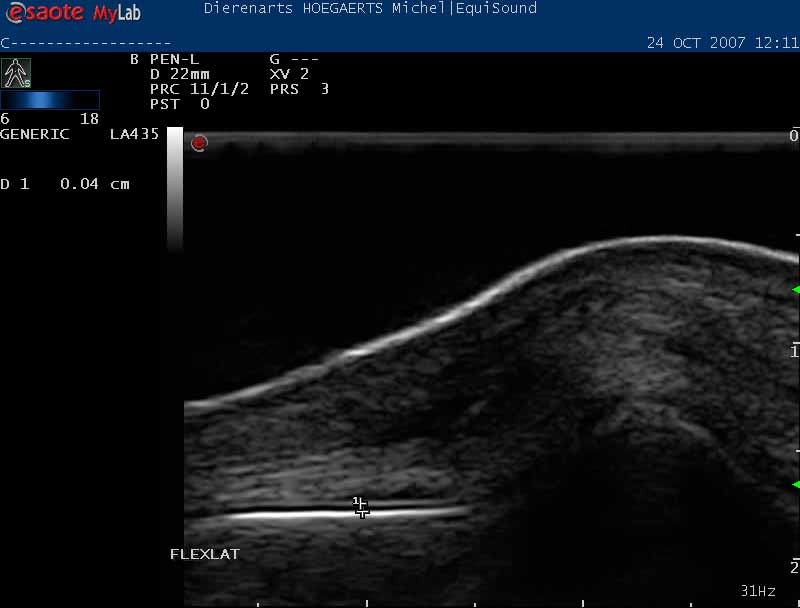
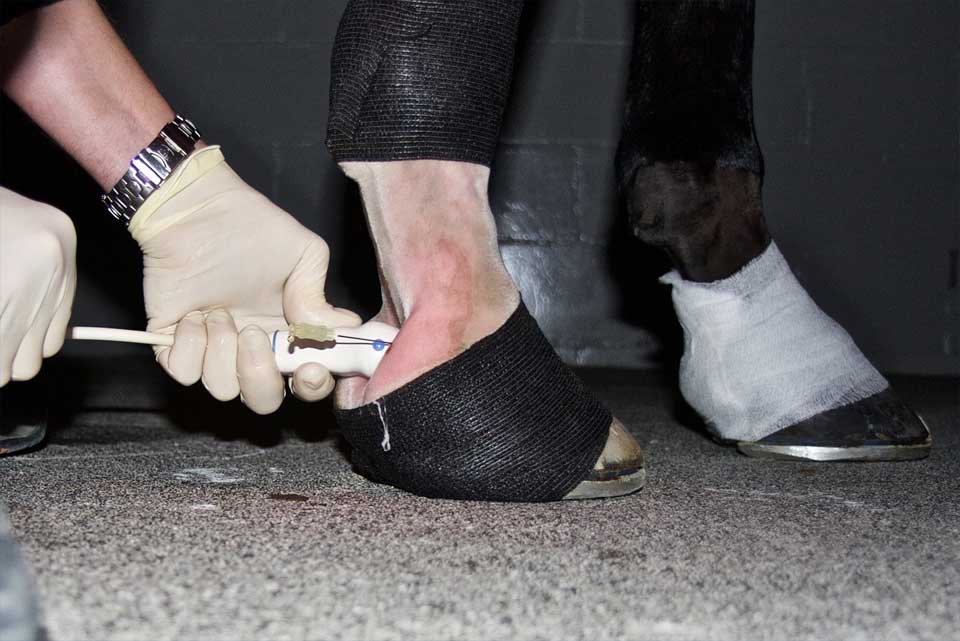
The examination of tendon injuries
Tendon injuries (tendinitis or tendinosis) of the extensor and flexor tendons of front and hind limbs are common.
Ultrasound examination remains the gold standard for diagnosing and monitoring tendon injuries. Performing an ultrasound is essential when a tendon is swollen, warm or painful to visualize the size and kind of the lesion. This way we can propose an adapted treatment and rehabilitation schedule. The goal is to optimize the repair tissue and to get the horse back to his previous training level as soon as possible.
Is your horse not performing as he should, or did you detect a swelling on a leg? Contact us and make an appointment at EquiSound. We also perform sports management, because a regular medical check-up can prevent (tendon) injuries.